|
|
|
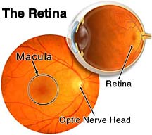
The retina is a very thin layer of tissue that lines the inner part of the eye. It is responsible for capturing
the light rays that enter the eye. Much like the film's role in photography. These light impulses are then sent
to the brain for processing, via the optic nerve.
|
|
|
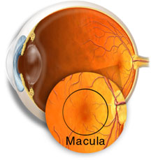
The macula is located roughly in the center of the retina, temporal to the optic nerve. It is a small and highly
sensitive part of the retina responsible for detailed central vision. The fovea is the very center of the macula. The
macula allows us to appreciate detail and perform tasks that require central vision such reading.
Copyright St. Luke's Cataract & Laser Institute
|
|
|
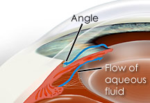
The aqueous is the thin, watery fluid that fills the space between the cornea and the iris (anterior chamber).
It is continually produced by the ciliary body, the part of the eye that lies just behind the iris. This fluid nourishes the
cornea and the lens and gives the front of the eye its form and shape.
Copyright St. Luke's Cataract & Laser Institute
|
I'm scheduled for Vitrectomy - membrane peel surgery on my right eye on 11/30/11 at 9:30 AM.
The vision out of my right eye reminds me of what it was like looking at the leaves on trees (when I was nearsighted) before
I got glasses when I was about 25 years old.
Click on the links below to learn more.
I have a condition named Epiretinal Membrane (Macular Pucker)
Here's an overview of what Epiretinal Membrane Pucker is.
Overview of Vitroctomy surgery
Animation of actual surgery
|